Esthetic Wax-up ,, Part 1
What is an Esthetic Wax-Up ?
An esthetic wax-up is a process in which wax is applied to a model of the patient’s teeth to simulate the procedure and results of planned reconstruction, repair, or enhancement of a smile.
Esthetic wax-ups are indicated when anterior restorations are planned to restore caries lesions, fractures, wear (attrition/erosion/abrasion), diastema, intrinsic discoloration, or microdontia that most commonly affect the lateral incisors (peg-shaped lateral) or to generally enhance a poor smile. Several factors should be considered before initiating the treatment, such as caries risk, periodontal health, existing restorations, alternative treatment options, and patient expectations.
First of all we have to put into consideration some principles that are very crucial to know before we start implementation of the procedure.

Dental Esthetics (Microesthetics)
Alignment of teeth
Properly aligned teeth are a major contributor in achieving the ideal smile. Tooth angulation from a lateral view represents the faciolingual alignment of teeth and determines the soft tissue support, lip prominence, and facial profile as well as the appearance of the teeth. For beautiful esthetics, the anterior teeth are facially inclined with their root apices toward the lingual (the maxillary teeth are slightly more inclined than the mandibular). If this angulation is decreased, the lips become less supported and therefore less esthetic, and the nose and chin will dominate the smile. On the other extreme, if the angulation is increased too much, this results in protruded incisors and increased incisal display, causing the smile to be unpleasant as well as increasing the chance of trauma to the maxillary incisors.
From the frontal view, the long axes of crowns are distally inclined, making the incisal portion mesial to the gingival portion of the crown. This is opposite the angulation of the root axis lines of these teeth, which tends to be mesially inclined, meaning that even though the entire tooth is angulated mesially as determined by the root axis line, the crown is angulated distally in relation to the root and to the midline of the tooth. Naturally, this distal inclination of the anterior crowns as seen from a frontal view should increase progressively from the central incisors to the canines (ie, it should be least noticeable with the central incisors).
Another component to be considered regarding the vertical alignment of anterior teeth is the relationship to the midline , which plays a major role in esthetics. A dental midline shifted from the facial midline by 2 mm or less may pass unnoticed. This is true as long as the central incisors are aligned vertically with their incisal plane parallel to the interpupillary line. Lack of proper vertical alignment of maxillary anterior teeth resulting in a canted midline is more esthetically unpleasant than a midline shift.

Morphology of anterior teeth
The morphologic components of anterior teeth that are apparent upon smiling include labial surfaces, incisal ridges, canine cusp tips and ridges, incisal embrasures, and interproximal contact areas.
Labial surfaces of anterior teeth are smooth and slightly convex in a mesiodistal and cervicoincisal direction with the height of contour occurring in the cervical third and extending approximately 0.5 mm labially. Anterior teeth are formed of four developmental lobes (growth centers). The lingual lobe is represented by the cingulum forming the height of contour at the lingual surface, and the three facial lobes are represented by the mamelons that appear in the newly erupted teeth and wear away with function but are not a feature of the adult permanent dentition. The middle lobe may be overpronounced, forming a slight elevation in the center of anterior teeth. In the canines, the middle lobe is more pronounced than in the incisors, resulting in their quite convex appearance. The labial surfaces are bounded by the mesiolabial and distolabial line angles, which are slightly rounded, with the distolabial line angle being more convex. As a result of the convexity in the center of anterior teeth produced by the middle lobe and the convex line angles at the borders, two shallow depressions appear mesial and distal to the middle lobe of the tooth.
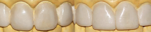
It is important to remember that the prominence of the middle lobe and hence the adjacent developmental depressions may not be apparent in every dentition, and in many cases the labial surface can be smooth between the line angle. In anterior restorations and esthetic wax-ups, these features are reproduced if they exist on the contralateral and adjacent teeth or whenever it is found more esthetically pleasing.
 |
| (a) Developmental depression in an extracted central incisor. (b) Esthetic wax-up with developmental depressions. |

Incisal edge position
The factors considered when positioning the incisal edges in smile design are degree of tooth display, phonetics, occlusion, relative tooth dimensions, and patient preference. The maxillary central incisal edges are the first to be located on an esthetic wax-up and provide a guide for the remainder of the anterior teeth. Ideally, they should be placed against the inner edge of the vermillion border at the wet-dry line of the lower lip when the patient pronounces the letter “F” and achieve tooth display of 2 to 4 mm at rest in an upright position in younger patients. In addition, contact between maxillary and mandibular incisal edges in protrusion should separate posterior teeth on the mounted casts. The maxillary lateral incisal edges are positioned 1 to 1.5 mm shorter than the central incisal edges.
 |
Canines have one strong prominent cusp. The distal cusp ridge is longer than the mesial cusp ridge in both maxillary and mandibular canines, making the cusp tip offset to the mesial side. The labial cusp ridge is quite prominent and runs on the labial surface of the canines, giving them a convex appearance in both mesiodistal and incisocervical directions. These features should be perfected in esthetic wax-ups for a natural appearance.
During an esthetic wax-up, the canine cusp tip position depends on a balance between the amount of tooth display as well as occlusion. Ideally the maxillary canine cusp tip falls on the smile arc on the same point as the maxillary central incisal edges and should allow the disocclusion of posterior teeth in lateral movement to achieve canine guidance.
Embrasures
Embrasures are the V-shaped spaces between the teeth that allow the escape of food, which essentially reduces food impaction and improves masticatory function. Incisal embrasures are located toward the incisal edge and terminate at the interproximal contact point. Their shapes and sizes are important in achieving natural esthetics and should be reproduced precisely in an esthetic wax-up.
For beautiful esthetics, incisal embrasures should become progressively larger posteriorly. Therefore, the embrasure is minimal between the two central incisors, larger between the central and lateral incisors, and largest between the lateral incisor and canine. This can be easily achieved during waxing by proper reproduction of the interproximal contact location and the incisal edge height and form as well as perfecting the point angles to the correct rounding. If these embrasures are made uniform, this results in an unnatural smile with interproximal contacts that are too long and produces a box-shaped appearance of teeth.

Interproximal contact area
Interproximal contact area is defined as the broad zone in which two adjacent teeth touch, bounded by the apex of the V-shaped gingival embrasure at the gingival end and the apex of the incisal embrasures at the incisal end.
Interproximal contact areas ideally follow the " 50:40:30 rule " in reference to the maxillary central incisor. Ideal proximal contact area between central incisors is about 50% of the length of the central incisors, between central and lateral incisors is about 40% the length of the central incisors, and between the lateral incisor and the canine is about 30% the length of the central incisors.
Interproximal contacts may influence dental esthetics by affecting the size of the embrasures as follows ; extending the interproximal contact areas gingivally decreases the size of the gingival embrasures and can help reduce or eliminate black triangles that may appear due to gingival recession, and extending the interproximal contacts further incisally reduces the size of the incisal embrasures. Interproximal contacts may also affect esthetics by creating an optical illusion whereby the teeth appear longer if the interproximal contact areas were increased and shorter if the interproximal contact areas were decreased.

Size of teeth/Tooth dimensions :
Maxillary lateral incisors are considered the playful part of the smile and are often asymmetric. They provide individuality and gender characterization by their curvatures. Straight line angles and relatively sharper incisal edges denote masculinity, while convex point and line angles with a round incisal edge denote femininity. Lateral incisors are the narrowest maxillary anterior teeth mesiodistally and their lengths are usually 1 to 1.5 mm shorter than the central incisors; therefore, they do not touch the smile arc and exhibit less wear with aging compared to the central incisors and canines due to diminished occlusal contact.
Maxillary canines play a critical point in creating a pleasing smile as they are the junction between the anterior and posterior dental segments and provide support to the corner of the mouth. Canines denote vigor and personality characterization. Masculine canines are aggressive with a sharper cusp tip, and feminine canines exhibit a more rounded cusp tip. For an esthetically pleasing smile, the canines should be the same length as the maxillary central incisors.

Establishing tooth width
A mathematical formula has been derived that calculates the width of the maxillary central incisor for any RED proportion given a fixed intercanine frontal view width. This width is determined by measuring the frontal view width between the distal aspects of the two maxillary canine teeth. The formula is as follows: (frontal view width of the 6 anterior teeth) / 2 (1 + RED + RED2) = width of central incisor.
Once the ideal width of the central incisor has been calculated, the width of the central incisor is multiplied by the desired RED proportion to determine the frontal view width of the lateral incisor. The resulting lateral incisor width is multiplied by the same RED proportion to yoeld the desired frontal view width of the canine.
The golden proportion could be defined as the 62% RED proportion, and is one of many RED proportions that can be applied. Thus, RED proportion has a more universal application.

The law of face
The face of a tooth is the area on the facial surface of anterior and posterior teeth that is bounded by the transitional line angles as viewed from the facial aspect. The transitional line angles mark the transition from the facial surface to the mesial, distal, cervical, and incisal surfaces. The tooth surface slopes lingually toward the mesial and distal surfaces and toward the cervical root surface from these line angles. Often no transitional line angle appears on the incisal portion of the facial surface; in this situation, the face is bounded by the incisal edge or the occlusal tip. Shadows are created as the light strikes the line angles, and the face of the tooth is the only area that reflects light. Therefore, the size of the face of the tooth determines the perceived size of the facial surface. Improper position or contour of transitional line angles will result in a wax-up or a restoration that appears dissimilar even if it is identical in size to the contralateral tooth because the light is being reflected from a smaller or larger surface.
Characterization
The purpose of smile design and esthetic wax-ups is to create a natural-looking, beautiful smile. However, the reality is that natural smiles are not necessarily perfectly symmetric, uniform in color, or perfect by scientific standards. In addition, perception of beauty is very subjective and differs between genders, ages, and cultures. Purely focusing on the scientific principles is not the best approach and may result in smiles appearing too uniform and perfect and thus perceived as fake. Achieving the ideal smile requires a combination of the esthetic principles and artistic creativity to match the individual’s personality.
Optical illusions
Some techniques may be used to make teeth appear narrower or wider if the space available was not equal to the ideal-sized teeth for esthetics. In addition to the techniques mentioned below, which can be incorporated in a wax-up, different stains and shades used in the porcelain can also provide the illusion of narrower or wider teeth.
 |
 |
| Rounding the point angles decreases the mesiodistal width of the incisal edges to make teeth appear narrower. Apparent width B is larger than A even though both teeth are equal in width. |
How to make teeth appear narrower in a wax-up :
How to make teeth appear wider in a wax-up :
 |
| Vertical characterization groove to make teeth narrower. |
 | ||||||
Horizontal characterization groove to increase the apparent width of teeth.
|
 |
| Making an esthetic wax-up appear narrower by moving the line angles toward the midline, rounding the point angles and incorporating vertical characterization lines. |
 |
| Moving the canine labial ridge distally in Fig. B to make the canine appear wider. |
 |
| Making the canine appear narrower and longer in Fig.B by moving the lavial ridge mesially, pointing the cusp tip and incorporating vertical characterization line. Stay tuned for " Part 2 " ,, How To Implement ? .. |
Islam Zakzouk
DSefa is a blog that's dedicated to bringing you high quality health, lifestyle tutorial and resources on different categories. It's main forus on user experience.


I guess the first 2 figures a & b are the same.
ReplyDeleteExcellent article as usual Dr.Eslam
thanks alot and sorry for the mistake , edited
DeleteThanks
ReplyDeleteyou're welcome
DeleteHelpfull
ReplyDeleteThanks dr.Islam
EID Mubarak
thank you so much dr Islam
ReplyDeleteIn the event that you are looking through on the web, at that point pay special mind to the ad in places like Kijiji, eBay or Craigslist, while in your nearby every day the notice ought to show up in the classifieds area.UV light sanitizer
ReplyDeleteExcellent article i love it
ReplyDeleteHi there, I discovered your blog per Google bit searching for such kinda educational advise moreover your inform beholds very remarkable for me.
ReplyDeletehttps://advance-esthetic.us/btl-aesthetics-vanquish
Excellent and simple article
ReplyDelete